-logo.png)


Fluorescence spectroscopy is a versatile technique frequently used for the analysis of the tertiary structure of proteins. Most proteins exhibit intrinsic fluorescence, predominantly derived from tryptophan and tyrosine residues. Changes in protein conformation caused by different solution conditions (pH, excipients, etc.), elevated temperature, storage and/or interaction with other biomacromolecules can be detected by means of fluorescence measurements. This is due to the fact that tryptophan fluorescence (selective excitation at 295 nm) is particularly sensitive to the local environment of the residue. Thus, any conformational change (e.g. unfolding, aggregation) leads to changes in fluorescence intensity and/or emission maximum. Apart from intrinsic fluorescence, the use of suitable fluorophores or dyes (such as Nile red, ANS, FITC, pyrene) is also available in order to probe protein conformational changes.
At the Theoretical and Physical Chemistry Institute (TPCI) of NHRF a variety of spectroscopic techniques is available. As far as fluorescence spectroscopy is concerned the spectrometer available through the Instruct-EL hub, is a time-resolved fluorescence system (NanoLog, Horiba-Jobin Yvon) with 3 detectors: steady-state & time-resolved fluorescence / 2D NIR photoluminescence map capability together with multiple laser lines and cell temperature control. Fluorescence spectroscopic services involve conformational studies of proteins (native structure), as well as the investigation of structural changes (unfolding, refolding, aggregation) caused by changes in solution conditions or interactions with ligands/macromolecules, either at steady state or time resolved and kinetics experiments. Moreover, the specific instrumentation includes a reflectance apparatus, suited for measurements of thin films and surfaces.

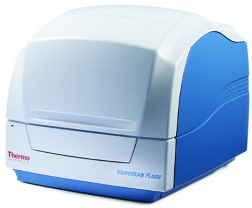
DESCRIPTION: Microplate reader Vis/UV/Fluorescence/ Luminescence
FUNDER: FP7-REGPOT-2009-1, No245866, "ARCADE"

DESCRIPTION: Protein structure determination and studies on physicochemical properties in protein aqueous solutions
FUNDER: EU-Capacities

DESCRIPTION: Fluorimeter equipped with temperature control system and polarizer accessory for fluorescence emission and anisotropy measurements
FUNDER:

DESCRIPTION: Fluorescence microplate reader for fluorescence emission and fluorescence polarization measurements equipped with heating and cooling unit, Fluorescence monochromator for top/bottom reading, Fluorescence variable bandwidth, Fluorescence polarization accessory, •Fluorescence dichroic mirrors
FUNDER: iNSPIRED
single cell fluorometer F2500 Hitachi (Karditsa)
The assay relies on the ability to quench the intrinsic fluorescence of tryptophan or tyrosine residues within a protein that results from changes in the local environment polarity experienced by the tryptophan(s) upon the addition of a binding partner or ligand. In other worlds the tryptophan or tyrosine should located in the binding pocket and thus interacts with the binding partner or ligand.
Fluorescence titration can determine affinity (Kd) and stoichiometry of a binding interaction (e.g. A + B ↔AB) in one experiment. It works by titrating reactant B into reactant A.
User should supply a sample usually purified protein (reactant A) and a ligand (reactant B)
Requirement
Reactant A
Purity protein > 95%
Concentration: 0.2 to 1μΜ
Volume: 1.5ml
Reactant B
Concentration: 50 to 500μM
Volume: 0.2ml
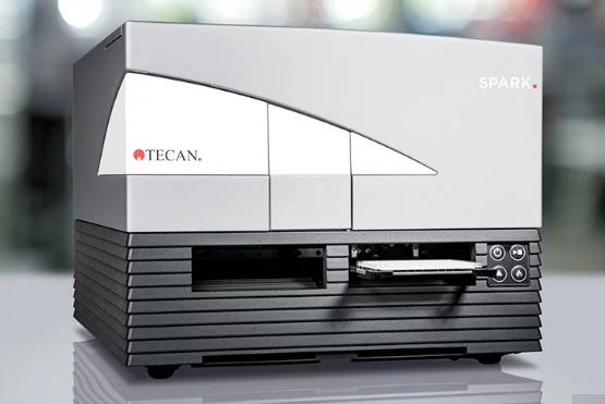
plate reader Karditsa F200 Tecan
fluorophore is excited with light that is linearly polarized by passing through an excitation polarizing filter; the polarized fluorescence is measured through an emission polarizer either parallel or perpendicular to the exciting light's plane of polarization.
Fluorescence titration can determine IC50 and Kd of a binding ligand to a protein
User should provide purified protein, fluorophore, ligand and one Black opaque 384‐well microplates (Corning)
Requirement
Purified protein
Purity > 80%
Concentration: 30-100 nΜ
Volume: 100 μL
fluorophore (tracer)
fluoroscine base tracer or other with absorption maximum at 494 nm and emission maximum of 512 nm and affinity (Kd) to protein 0.01 to 1 μM
Concentration: 100-1000 nΜ
Volume: 100 μL
ligand
Concentration: 100-1000 nΜ
Volume: 200 μL

DESCRIPTION:
FUNDER:

DESCRIPTION:
FUNDER:

DESCRIPTION:
FUNDER: INSPIRED-RIS
