

UV-Vis spectroscopic measurements consist one of the most widely used methods for protein quantification. This is due to the fact that the tryptophan and tyrosine (and to a lesser extent the phenylalanine) residues of proteins strongly absorb at 280 nm. Hence, the corresponding absorbance is used for the calculation of protein concentration, based on specific quantification formulas. If the protein sample does not have tryptophan or tyrosine, the concentration can still be easily measured by the absorption at 205 nm in which the peptide bonds are analysed directly (Scopes method). Conformational studies can also be performed under special conditions.
The spectroscopic equipment of TPCI/NHRF accessible through the Instruct-EL hub includes a UV-Vis-NIR spectrophotometer (Lambda 19 by Perkin Elmer), which is a general-purpose instrument for absorption spectroscopy in the ultraviolet, visible, and near infrared spectral regions. It allows measurement of transmittance and reflectance of diffuse or scattering materials as a function of wavelength. Moreover, the optical scheme consists of a double beam double monochromator configuration, while the useable wavelength range is 250 – 2500 nm with temperature control. UV-Vis spectroscopic services are mainly focused towards protein quantification and concentration determination. Moreover, reflectance measurements of thin films and surfaces is also available.
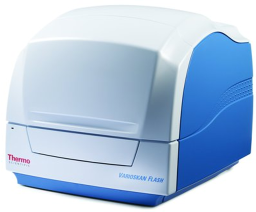
A double beam UV-Vis spectrophotometer (HITACHI U-2001) is available in the HPI’s Bacteriology laboratory measuring absorbance at the range 190 – 1100 nm with temperatures being controlled trough a water circulation apparatus. The device can be used for a variety of applications such as protein and nucleic acid quantification, wave length scans, and time scans permitting the real time monitoring of enzymatic reactions for steady state kinetic analyses and inhibition studies. The instrument is PC controlled and the relevant analyses can be performed with the UV Visions software (Hitachi).
Visit the provider's website: http://inspired.aua.gr/instruct/
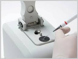
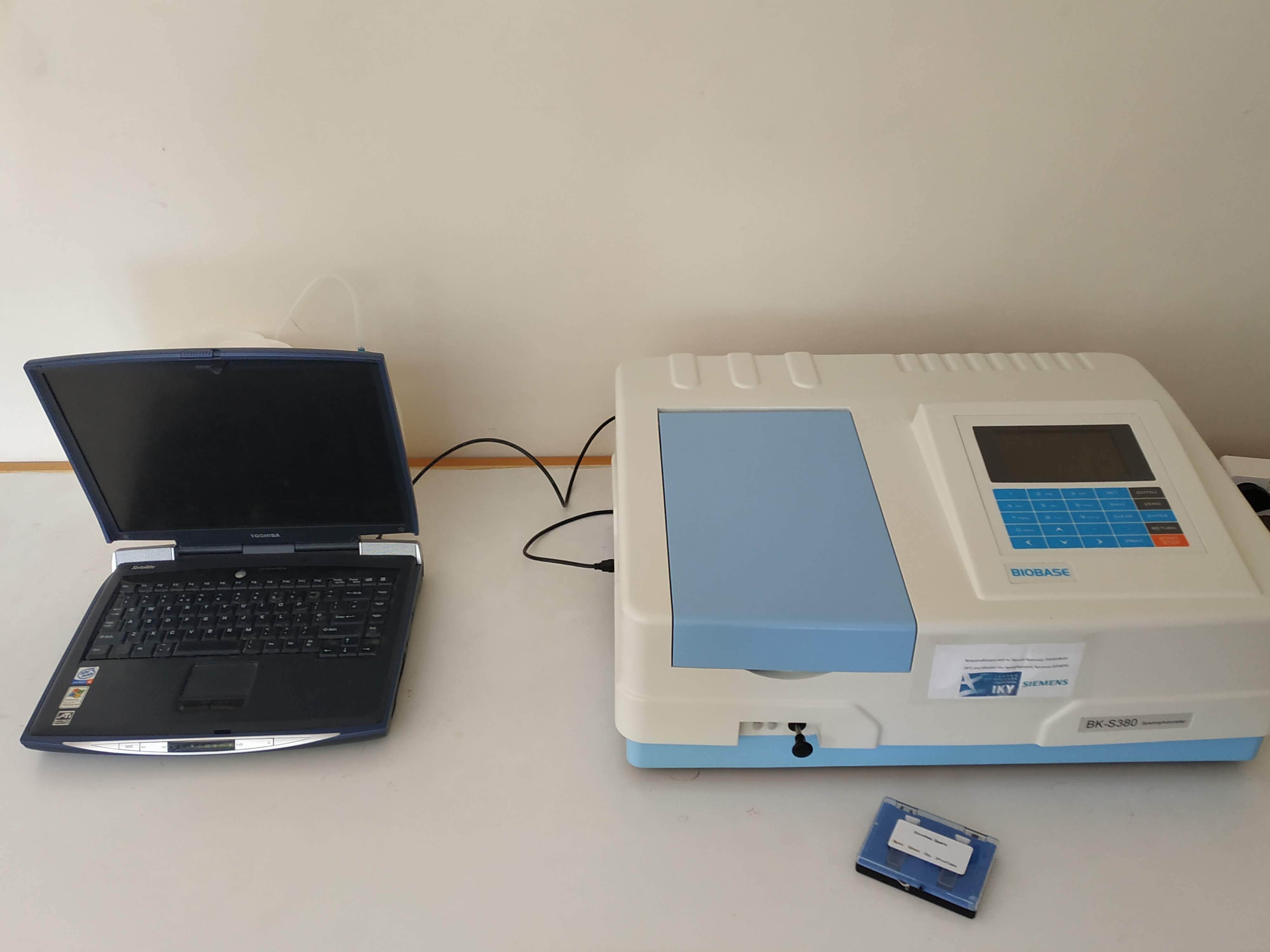
Basic Functions:
Photometry: Test Abs., Transmittance and Energy by fixed wavelength
Quantitative: Linear fit and Linear fit through zero two modes
Coefficient, Standard Sample input and Standard Sample read three modes to establish standard curve
Establish A=K1*C+K0, can search original data, graph curve, parameters settings
Can save 240 group curves, can test 240 data in each curve
Double wavelength, Triple wavelength test functions
Kinetics: Used for time course scanning or reaction rate calculations△A/t, can search all data.
Multi-Wavelength: Can test Transmittance and Abs. with 8 different wavelengths at most
Scanning: User can set the scan range from 190nm to 1100nm to test the max. Abs. peak value,
can do derivation, arithmetical operations to the graph.
Biology: DNA/Protein, (Optional: UV, Lowry, BCA, CBB and Biuret)
The Hitachi's U-5100 UV-Visible Spectrophotometer is a general-purpose instrument for absorption spectroscopy in the ultraviolet, visible, and near infrared spectral regions. It delivers a compact, lightweight package with remarkable power savings and a long life for its light source. The optical scheme consists of a Seya-Namioka mount monochromator configuration and the useable wavelength range is 190-1100 nm with temperature control. UV-Vis spectroscopic services are mainly focused towards protein quantification and concentration determination. Moreover, It contains automatic switching of 6 cells by using an automatic 6-cell turret and a high-speed scanning (12,000 nm/min) deliver a substantial reduction in measurement time (approximately 60% reduction).
This is some info
Dr. George Mousdis (born 1962, Diploma in Chemistry NKUA 1986, Ph.D NKUA 1990) is Director of Research at TPCI-NHRF. His current research interests lie in the synthesis of hybrid low dimensional materials, electronic spectroscopy, sol-gel chemistry, fluorometry and sensors. Author or coauthor in 115 publications in refereed journals, 22 publications in conference proceedings or book chapters and numerous announcements at significant international scientific conferences. His work has more than 1570 citations (h index=20). He coordinated or participated in more than seventeen national and international research projects. He is a regular referee for international journals in the fields of physical chemistry and materials science and reviewer for EU, international and national research proposals and projects.
Professor Elias Eliopoulos is the Director of the Genetics Laboratory at the Biotechnology Department of the Agricultural University of Athens (A.U.A.) and leads the bioinformatics and structural biology group. He is professor of Biochemistry, a biophysicist and crystallographer by training with considerable experience in biomolecular structure analysis, epitope mapping of protein receptors, genetic polymorphisms and variation, cloning, with experimental and computational methods, protein structure prediction, ligand and drug design, protein design, in silico antibody design and biosoftware development. The group has experience and international reputation on computational protein folding, ab initio and homology modeling of proteins derived from edge gene research and membrane protein modeling. The group is also involved in many ligand computational design projects for pharmaceutical, medicinal and agricultural applications.
Basic Functions:
Photometry: Test Abs., Transmittance and Energy by fixed wavelength
Quantitative: Linear fit and Linear fit through zero two modes
Coefficient, Standard Sample input and Standard Sample read three modes to establish standard curve
Establish A=K1*C+K0, can search original data, graph curve, parameters settings
Can save 240 group curves, can test 240 data in each curve
Double wavelength, Triple wavelength test functions
Kinetics: Used for time course scanning or reaction rate calculations△A/t, can search all data.
Multi-Wavelength: Can test Transmittance and Abs. with 8 different wavelengths at most
Scanning: User can set the scan range from 190nm to 1100nm to test the max. Abs. peak value,
can do derivation, arithmetical operations to the graph.
Biology: DNA/Protein, (Optional: UV, Lowry, BCA, CBB and Biuret)
Dr. Stathis Frillingos is Professor of Biological Chemistry at the University of Ioannina, Department of Medicine, and Director of the Interinstitutional Interdepartmental Programme of Postgraduate Studies in Molecular and Cellular Biology and Biotechnology at the University of Ioannina, Greece (http://msc-mcbb.ac.uoi.gr). His research team focuses on the analysis of structure-function-specificity and evolutionary relationships of membrane transporters, with current emphasis on Solute Carriers responsible for the transmembrane transport of nucleosides, nucleobases and their antimetabolite analogs. His research studies include the functional characterization of new nucleobase/nucleoside transporters from microorganisms and analysis of their binding-site interactions with inhibitors and antimetabolite compounds as potential antimicrobial drugs (http://www.frillingoslab.gr).
EDUCATION
2019: PhD in Structural Biology and Protein Biomaterials (UoC, IMBB-FORTH - Greece)
2012: M.Sc. in Protein Biotechnology (UoC-Greece)
2009: Diploma Degree in Biology (UoC-Greece)
WORKING EXPERIENCE
Oct 2019 – Now: Post-Doctoral Researcher at I.M.B.B.-F.O.R.TH, Greece
(Laboratory of Structural Biology)
Sep 2020 – Now: Autonomous teaching and exam organizing of the under-graduate courses
"Enzymatic Biotechnology" and “Protein Engineering” at the Dept. of
Biology of the Uni. of Crete
2014 – 2020: Experienced scientist position at I.M.B.B.-F.O.R.TH, Greece (InnovCrete project /FP7-Regpot program) and at University of Crete
Oct. 2010 –Jan. 2011: Laboratory Teaching Assistant of Microbiology Teaching Lab
at the Dept. of Biology, Uni. of Crete
SELECTED PUBLICATIONS
*Kefala K., Kotsifaki D., Providaki M., Kapetaniou E., Rahme L. and Kokkinidis M. Purification, crystallization and preliminary X-ray diffraction analysis of the C-terminal fragment of the MvfR protein of Pseudomonas aeruginosa. (2012). Acta. Cryst F, 68, 695-697
*Kefala A., Kotsifaki D., Providaki M., Amprazi M. and Kokkinidis M. Expression, purification and crystallization of a protein resulting from the inversion of the aminoacid sequence of a helical bundle. (2017). ). Acta. Cryst F, 73, 51-53
Dimovasili C., Fadouloglou V.E., Kefala A., Providaki M., Kotsifaki D, Sarrou I., Plaitakis A., Zaganas I. and Kokkinidis M. Crystal structure of glutamate dehydrogenase 2, a positively selected novel human enzyme involved in brain biology and cancer pathophysiology. Journal of Neurochemistry (accepted)
RESEARCH PROFILE
Being in a Structural Biology group since 2011, Dr. Kefala A. has studied deeply the protein folding problem, elucidating the relation between amino acid sequence and structural properties of α-helical bundles. The knowledge obtained was used for protein design leading to bio-derived materials for bio-medical applications and basic research through structural studies of protein folding through reverse sequences.
Notable achievements include:
The elucidation of the protein folding of mutants of the α-helical protein Rop that is used as a canonical paradigm of this structural motif.
Protein design of novel protein-based biomaterials with specific engineered properties.